


FDG PET scan: an advanced tool in disease diagnosis and management
PET scan (Positron Emission Tomography) is a medical imaging technique that is particularly useful in the diagnosis and management of cancer, heart and neurological diseases. This method uses radioisotopes to provide detailed information about the metabolic activity of tissues. One of the most widely used radioisotopes in PET scans is fluorodeoxyglucose (FDG), which is specifically designed to examine glucose metabolism in cells.
How FDG PET scans work
FDG is a radioactively labeled glucose analog. After FDG is injected into a patient, it is taken up by metabolically active cells. Cancer cells typically use more glucose than normal cells, so FDG will accumulate more in cancerous areas. After a certain amount of time, images of the patient’s body are taken using a PET scanner that show the distribution of FDG.
Clinical applications
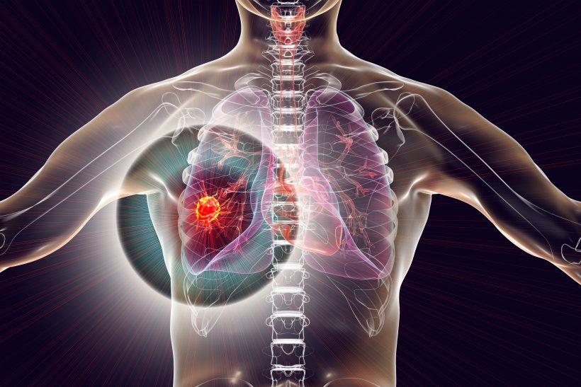
1. Cancer diagnosis: FDG PET scans are widely used to detect and stage various types of cancers, such as breast, lung, and lymphoma.
2. Treatment evaluation: This method can be used to assess patients’ response to chemotherapy or radiation therapy.
3. Heart disease diagnosis: FDG PET scans are also used to evaluate heart function and identify areas damaged by a heart attack.
4. Neurological diseases: This technique is also used in the diagnosis of diseases such as Alzheimer’s and Parkinson’s.
Advantages and disadvantages
Advantages:
– High accuracy in identifying metabolically active areas.
– Ability to provide information about the biological activity of tissues.
Disadvantages:
– High cost of testing.
– Requires specialized equipment and experienced medical staff.
– Side effects from radioactive radiation (although usually low).
Conclusion
FDG PET scanning is a valuable tool in modern medicine that has revolutionized the diagnosis and management of diseases. Given its high accuracy and ability to provide comprehensive information about the body’s metabolic status, this method remains one of the most important techniques in the field of medical imaging. Further research is ongoing to increase accuracy and reduce associated costs so that it can be more accessible to patients.